SPONGES
Organized as a loose aggregation of various independent cells rather than via distinct tissue types, a sponge is essentially a water filtration device. Siliceous sponges (PO9–12) are an exception in that these cells are multi-nucleate (many central bodies with genetic material) without cell boundaries.
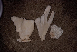
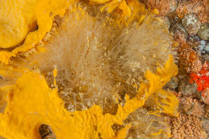
Central to this is a system of pores, canals and chambers, often with a complicated layout and lined with collar cells or collar bodies in one special group of sponges. These flagellate cells or bodies—each with a single hair-like structure—move seawater through the sponge while removing microscopic planktonic food. Sponges typically are supported by a skeletal framework of fibres often accompanied by glass- or chalk-like slivers called spicules. Photograph A shows the spicule structure of three dried specimens. Photograph B features a living sponge with its spicule skeleton exposed.
Sponge classification and recognition is a most problematic and frustrating subject for a naturalist. Indeed, academic professionals who spend their careers studying the group are likewise challenged—a fact that leads to much uncertainty in scientifically oriented sponge publications. Historical inconsistencies and funding limitations overlay the fundamental difficulty of studying sponge anatomy. As such, sponge taxonomy is rendered a “permanent work in progress.”
One approach to species identification of sponges is via their shapes, a standard used for most groups of organisms. This is workable in many instances, but numerous sponge species have few large, obvious diagnostic features. This is particularly true of encrusting forms that grow in mat-like carpets over whatever shape and type of surface is available. Environmental influences often dictate sponge growth patterns, further complicating this anatomical approach.
When applied with shape, colour can sometimes be an aid to sponge identification but must be considered only a factor of partial and limited value.
Traditional considerations of the aforementioned issues have led experts to focus on the “micro-anatomy” of sponges—namely the organization and types of fibres and/or spicules. The size and shape of these building blocks and their combinations have proven significant for identifying most species. Unfortunately, such precise determination involves microscopic examination of spicules, a task that most often must be preceded by sampling and preparation. Once spicule terminology is understood, complicated “keys”—jargon for intensive road maps—must be carefully applied. Even professional “spongophiles” have difficulty with such intricacies. As a result, numerous “forms” remain inadequately known, even nameless and essentially unclassified. Obviously this mitigates against successful identification of sponges by the beach-side naturalist or underwater explorer!
During preparation of this portion of the book, we became as confused as nearly everyone else about sponges and sought out Dr. Bill Austin of the Marine Ecology Centre in Sidney, BC—the acknowledged Pacific Northwest sponge guru. His invaluable assistance is greatly appreciated. Fundamental to his advice was that the understanding of sponges is in a state of flux and that caution should be exercised with respect to identification, particularly when samples are unavailable for inspection—as is the case for some entries presented here. When relevant, we mention such possibilities within a “species” account. The identification of some sponges that were photographed but not sampled proved hopeless, and a “best guess” would prove counterproductive. These sponges are presented at the end of this section.
The estimate of at least 260 marine species in the Pacific Northwest, made at the time of original publication, has proved to be very conservative. Consequently, the section labelled “Undetermined Sponges – A selection” has been adjusted and numerous entries now follow PO85/86 with a brief preamble.